|
There are no efficient lenses for
X-rays and therefore no X-ray microscopes. Still, there are ways
to image defects with X-rays. |
|
 |
The essential part for imaging
defects in crystals is the diffraction
of the X-rays in the crystal lattice. This is in contrast to the
conventional X-ray imaging technique in medical applications were the
differential absorption of X-rays in
differently dense tissue is used. |
|
The basic principle (which is also
valid for imaging with electron beams in the transmission electron microscope)
is shown below: |
|
|
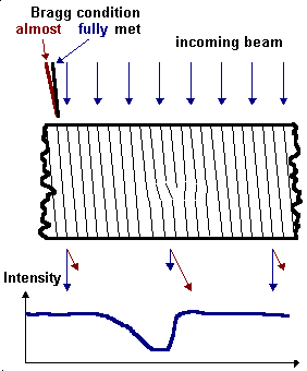
The specimen is oriented with respect
to the incoming wave in such a way that the Bragg-condition for diffraction is only met (or
nearly met) for just one set of lattice
planes. |
|
|
All defects with strain fields will
locally deform the lattice and thus change the Bragg condition locally. The
intensity of the diffracted beam will react to this and vary around defects.
This is schematically shown below |
|
|
|
|
|
|
|
In the example, the specimen is oriented in such
a way that the Bragg condition in the perfect part of the crystal is almost,
but not quite met. There will be no diffraction or, more quantitatively
speaking, a rather low intensity of the diffracted beam. The primary beam thus
is transmitted almost without any losses. |
|
To the left-hand side of the edge dislocation,
the strain field bends the lattice plane locally into the Bragg position. In
this area the primary beam is strongly diffracted and loses intensity. |
|
|
|
The intensity of the diffracted beam is mirror
symmetric to the primary beam. |
|
|
|
|
|
|
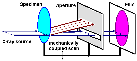
For the imaging of defects (typically
in Si-wafers, with or without processing) the following basic set-up is
used. |
|
|
|
|
|
|
|
|
|
An X-ray source with a thin
"one-dimensional" beam cross-section illuminates a line of the wafer.
Only the primary beam (or, for dark-field imaging, the diffracted beam) is
admitted through an aperture on the film. Wafer, aperture, and film are scanned
through the beam. |
|
Some examples of X-ray
topography are given in the following links;
another one we have already
encountered before. |
|
|
Total view and resolution limit |
|
|
Case study in bipolar technique |
|
The strengths and weaknesses of
X-ray topography are quite apparent: |
|
|
|
|
| Strength |
Weaknesses |
- Imaging of large wafers with good resolution (ca. 5 µm)
possible
- Detailed analysis (e.g. Burgers vectors) possible within limits
- No specimen preparation necessary
|
- Very expensive
- rather long exposure times even with powerful (typically 50 kW)
X-ray tubes
- Resolution/sensitivity not good enough for single/small defects
|
|
|
|
|
© H. Föll